New Fluorescent Probe Enhances Imaging of Nonalcoholic Fatty Liver Disease
A novel cholesterol-biomimetic fluorescent probe significantly improves the imaging of nonalcoholic fatty liver disease, offering a non-invasive diagnostic alternative.
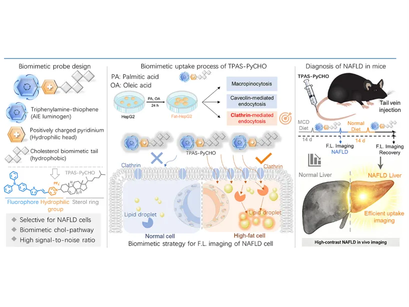
The TPAS-PyCHO+ probe shows a 4.81-fold increase in fluorescence in steatotic cells.
It selectively targets hepatocytes via the NPC1L1-mediated endocytosis pathway.
In NAFLD mice, the probe demonstrated a liver fluorescence peak of 2.28 times the initial intensity.
A groundbreaking fluorescent probe designed to mimic cholesterol has been developed to enhance imaging of nonalcoholic fatty liver disease (NAFLD). Researchers from Beijing University of Chemical Technology introduced TPAS-PyCHO+, which utilizes a unique mechanism to selectively target diseased liver cells. This innovative approach aims to address the limitations of traditional diagnostic methods, which often involve invasive biopsies and lack sensitivity for early-stage conditions.
NAFLD is recognized as the most prevalent chronic liver condition globally, with current diagnostic techniques like ultrasound and MRI falling short in detecting mild steatosis. The introduction of fluorescent probes has emerged as a promising alternative; however, existing options have struggled with specificity. The newly developed TPAS-PyCHO+ probe leverages the hyperactive cholesterol uptake pathway in steatotic hepatocytes, allowing for improved imaging accuracy.
The TPAS-PyCHO+ probe operates by mimicking the structure of cholesterol, featuring a triphenylamine-thiophene unit and a positively charged pyridinium salt. In laboratory tests, the probe exhibited a remarkable 4.81-fold increase in fluorescence in steatotic HepG2 cells, surpassing the performance of existing commercial dyes. Furthermore, it demonstrated a high degree of colocalization with other lipid droplet dyes, indicating its effectiveness in targeting pathological lipid accumulation.
The implications of this research extend beyond mere imaging capabilities. The ability to selectively visualize NAFLD could lead to earlier diagnoses and better monitoring of disease progression. In animal studies, the probe showed significant liver accumulation in NAFLD mice, with fluorescence levels peaking at 2.28 times the initial intensity compared to healthy controls. This suggests that TPAS-PyCHO+ could serve as a valuable tool for assessing liver health in both clinical and research settings.
Looking ahead, the researchers believe that the principles behind TPAS-PyCHO+ could be adapted for imaging other metabolic diseases characterized by similar endocytic dysfunctions. As the scientific community continues to explore non-invasive diagnostic methods, this cholesterol-biomimetic probe represents a significant advancement in the field of liver disease imaging.