New Study Reveals Enhanced CT Techniques for Temporal Bone Lesions
A recent study demonstrates that combining subtraction iodine maps with deep learning reconstruction significantly improves CT imaging for temporal bone lesions.
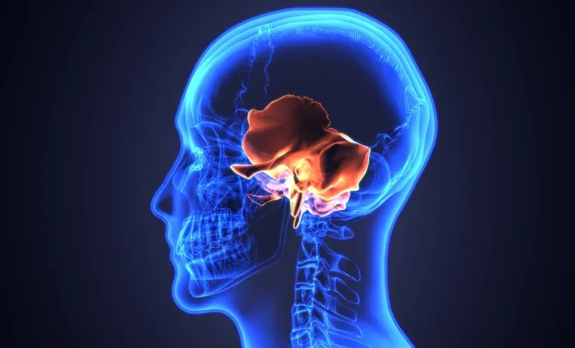
Combination of subtraction iodine maps and deep learning enhances CT image quality.
Study involved 77 patients with notable improvements in high contrast lesions.
Findings suggest potential for better pre-surgical assessment and treatment planning.
A recent prospective study has highlighted the effectiveness of combining subtraction iodine maps (SIM) with deep learning reconstruction (DLR) in improving the assessment of temporal bone lesions through computed tomography (CT). This innovative approach was found to significantly enhance image quality and the visualization of bone involvement in patients, particularly those with lesions exhibiting high contrast enhancement.
The study evaluated 77 patients who underwent enhanced temporal bone CT scans. Researchers compared four imaging techniques: conventional enhanced CT using hybrid iterative reconstruction (HIR), enhanced CT with DLR, SIM combined with HIR, and SIM combined with DLR. The results indicated that the SIM-DLR method outperformed conventional techniques, especially in cases where lesions showed pronounced contrast enhancement.
Two radiologists independently assessed the images based on overall quality, confidence in detecting lesions, delineation of contours, and visualization of temporal bone involvement. The study also included quantitative measures such as image noise and contrast-to-noise ratio (CNR). The findings revealed that the SIM-DLR technique significantly improved scores across all qualitative measures while also reducing image noise and enhancing lesion CNR.
The implications of this study are substantial for clinical practice. By improving the quality of CT images, particularly for hypervascular lesions, clinicians may be better equipped to assess the extent of disease prior to surgical interventions and to plan appropriate treatment strategies. However, the research was limited to a single center with a relatively small patient cohort, which may affect the generalizability of the results.
Looking ahead, the researchers advocate for larger, multicenter studies to further validate the clinical reliability and applicability of the SIM-DLR technique in assessing temporal bone lesions. Such studies could pave the way for broader adoption of these advanced imaging techniques in clinical settings.