Elastography Enhances Liver Fibrosis Detection in Thalassaemia Patients
Recent research highlights the effectiveness of transient elastography combined with biomarker scores in identifying liver fibrosis in thalassaemia.
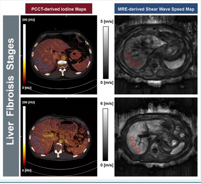
Transient elastography shows 85% sensitivity for detecting liver fibrosis in thalassaemia patients.
The method is less invasive than traditional liver biopsy, making it suitable for regular monitoring.
Further research is needed to validate these findings and improve diagnostic accuracy.
A recent systematic review has revealed that transient elastography (TE), when paired with non-invasive biomarker scores, can significantly enhance the detection and staging of liver fibrosis in individuals suffering from thalassaemia. This development is crucial as patients with thalassaemia frequently experience liver complications due to iron overload from blood transfusions and increased iron absorption, making effective monitoring essential.
Historically, liver biopsy has been considered the gold standard for assessing liver fibrosis; however, its invasive nature limits its practicality for ongoing evaluations. The systematic review analyzed data from nine studies involving 500 participants and focused on 12 diagnostic strategies, with TE being the only imaging technique assessed. The findings underscored TE's potential, particularly when used alongside the Fibrosis-4 (FIB-4) score, achieving an impressive sensitivity of 85% for identifying fibrosis at METAVIR stage F1 or higher.
In addition to detecting early fibrosis, TE also demonstrated promise in identifying cirrhosis. Researchers noted that by utilizing cut-off values between 11 and 13 kPa and integrating TE with other scores like APRI or FIB-4, diagnostic accuracy for METAVIR F4 disease improved significantly. This non-invasive approach could greatly benefit thalassaemia patients, who often require frequent monitoring but are deterred by the risks associated with liver biopsy.
Despite the encouraging results, the review's authors caution that the current evidence base is limited. Most diagnostic tests were assessed in only a few studies per fibrosis stage, and variations in reference standards hindered the ability to pool data effectively. They emphasized the necessity for additional research to validate these findings and to establish a consistent reference standard for liver biopsy in future studies.
As research in this area continues to evolve, the integration of non-invasive techniques like TE into routine monitoring could revolutionize the management of liver health in thalassaemia patients, making it more accessible and less invasive, ultimately improving patient outcomes.